Reprint from 3dprint.com

While most of us have either been the patient undergoing a surgical procedure or a loved one waiting and worrying, few of us have had to be on the other side as the trained surgeon, who often bears the responsibility of performing numerous delicate procedures with lives in his hands on a daily basis. Many of us are thrilled to hear about the innovations 3D printing offers to the medical world as people are saved with progressive implants and devices and quality of life is improved. 3D models are becoming more and more common not only for educating the patient on what is happening, but also for guiding surgeons in diagnosis as well as in treatment and the surgical process. Certain procedures may be completely new for a surgeon and while the 3D model is what he uses to study and practice on, it is probably the same 3D printed model that goes into the operating room with him and potentially shaves hours off the procedure, as well as plenty of stress.

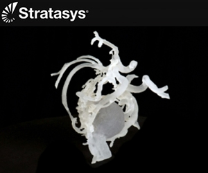
Vein of Galen malformation displayed in 3D model [Credit: Boston Children’s Hospital]

Vein of Galen malformation displayed in 3D model [Credit: Boston Children’s Hospital]

Recently, at Boston Children’s Hospital, surgeons were looking toward several tricky pediatric procedures. Thanks to 3D printing, they were able to fabricate 3D models of the anatomies of four children’s brains and not only examine what to do, but they were then able to rehearse for as much time as was required. Stricken with cerebrovascular malformations by way of abnormalities in their brain’s blood vessels, all four kids were considered to be high-risk cases. “These children had unique anatomy with deep vessels that were very tricky to operate on,” says Boston Children’s neurosurgeon Edward Smith, MD, co-director of the hospital’s Cerebrovascular Surgery and Interventions Center. “The 3D-printed models allowed us to rehearse the cases beforehand and reduce operative risk as much as we could.” The youngest patient was a mere two months old, and was suffering from a rare affliction called a vein of Galen malformation. This is a condition where instead of connecting with capillaries, arteries are connected directly to veins. A special technique was required to seal off the blood vessels internally. “Even for a radiologist who is comfortable working with and extrapolating from images on the computer to the patient, turning over a 3D model in your hand is transformative,” says Darren Orbach, MD, PhD, Chief of Interventional and Neurointerventional Radiology at Boston Children’s and co-director of the Cerebrovascular Surgery and Interventions Center. “Our brains work in three dimensions, and treatment planning with a printed model takes on an intuitive feel that it cannot otherwise have.” As they reported in a paper published in the Journal of Neurosurgery: Pediatrics, of which Dr. Smith is the senior author, a 3D resin printer was used to make a 3D model that basically put the children’s brains in the surgeons’ hands for inspection. The data for the models was created with magnetic resonance and MR arteriography data. There they could see, in detail, the vessel malformations, along with nearby normal blood vessels. They were also able to 3D print peripheral areas as needed also. In measuring the models, the doctors were able to see that they were a stunning 98% accurate in comparison to their actual brains.

A 3D printed model displaying AVM [Credit: Boston Children’s Hospital]

A 3D printed model displaying AVM [Credit: Boston Children’s Hospital]

Three of the four children, ranging up to the age of 16, had arteriovenous malformations which involves tangles of arteries and veins. All of the surgeries were successful and resulted in at least a 12%, or 30-minute, reduction in surgery time. That may not sound like a lot of time out of your day, but for kids under anesthesia, having the procedure reduced by just that simple amount could make a dramatic difference. “3D printing has become a regular part of our process,” says Dr. Smith. “It’s also a tool that allows us to educate our junior colleagues and trainees in a way that’s safe, without putting a child at risk.”

hospital

As is common in hospitals after using 3D printing for one of these major procedures, the surgeons plan to continue constructing 3D printed models as needed. While they may not need to draw on the equipment on a daily basis, certainly it gives surgeons a new level of enthusiasm and peace of mind regarding new procedures that are required. With the ability to take a better look, in 3D, and the ability to practice until they have it down, everyone benefits all around. It’s also in cases like these that everyone from patients and their families to many different members of medical staff and doctors become acquainted with 3D printing–and its wonders–when previously they were not. Discuss this latest use of 3D printed models for surgeries in the Surgeons 3D Print Models of Children’s Brains forum thread over at 3DPB.com. According to Boston Children’s Hospital, SIMPeds director Peter Weinstock, MD, PhD, was first author on the paper; co-authors were Orbach, Sanjay Prabhu, MBBS, FRCR, and Katie Flynn, BS, ME, all of Boston Children’s Hospital. The study was supported by the Lucas Warner AVM Research Fund and The Kids At Heart Neurosurgery Research Fund.